Younger-Looking Brains Linked To Better VNS Outcomes
Source: Brain stimulation
Summary
What was studied
Researchers built a "brain-age" tool from structural MRI scans. They trained it on scans from 2,623 healthy people ages 1.9 to 90 years to estimate how old a brain looks based on its structure, using a model designed to capture nonlinear developmental patterns. They evaluated how accurate the tool was with cross-validation and in an independent pediatric cohort.
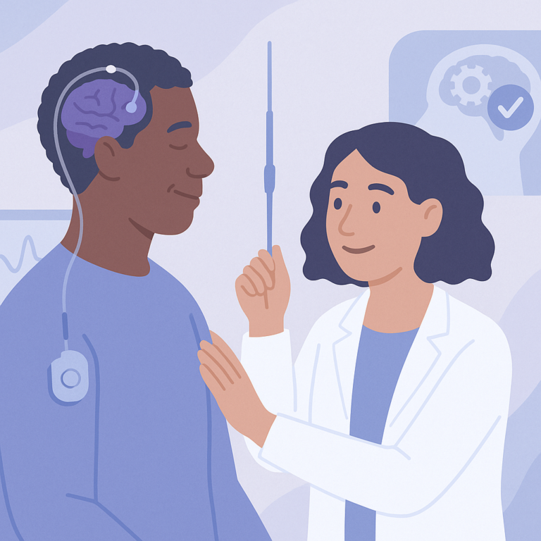
Next, they applied this tool to a multicenter group of 126 children with drug-resistant epilepsy who were treated with vagus nerve stimulation (VNS). They compared each child's estimated brain age with their actual age to calculate a "brain-age gap," then examined whether that gap related to seizure burden before surgery and seizure outcomes after VNS.
What they found
The brain-age model was reported to be accurate and to generalize well to an independent pediatric cohort. In the children who received VNS, brain-age gap was significantly higher than in age- and sex-matched controls, suggesting greater structural divergence from typical age-referenced patterns. Brain-age gap correlated with baseline seizure burden and with postoperative changes in seizure frequency. Children with a lower brain-age gap before surgery had a significantly higher likelihood of being clinical responders to VNS. Brain regions contributing disproportionately to these differences included the cingulate cortex, thalamus, nucleus accumbens, and prefrontal cortex, which align with VNS-related circuitry.
Limits of the evidence
This study does not prove that brain-age gap causes better or worse VNS outcomes; it reports associations. The VNS group included 126 children, and the abstract does not report how well the measure predicts outcomes for an individual child in clinical practice. The abstract also does not provide details on seizure types, other treatments, MRI differences across centers, or duration of follow-up. Because only the abstract is available here, important methods and possible sources of bias are not fully described.
For families and caregivers
This study suggests that an MRI-based measure of brain development might help support prognosis for children being considered for VNS. That could be useful when families and clinicians are discussing treatment options for drug-resistant epilepsy. But it is not a proven stand-alone decision tool, and it should not be seen as a yes-or-no test for whether VNS will work.
What to watch next
Useful next steps would include larger studies to test how well this MRI measure helps predict VNS response for individual children across different clinical settings.
Terms in this summary
- drug-resistant epilepsy
- Epilepsy in which seizures continue despite trying appropriate seizure medicines.
- vagus nerve stimulation (VNS)
- A treatment that sends regular electrical signals to the vagus nerve, usually through a device implanted in the chest, to help reduce seizures.
- structural MRI
- A brain scan that shows the brain's anatomy and shape.
- brain age
- An estimate of how old a brain looks based on scan features, which may differ from a person's actual age.
- brain-age gap
- The difference between estimated brain age and actual age.
- clinical responder
- A patient who has a meaningful improvement after treatment after VNS, as defined by the study.
- biomarker
- A measurable sign in the body or on a test that may help reflect disease burden or treatment response.
Free: Seizure First Aid Quick Guide (PDF)
Plus one plain-language weekly digest of new epilepsy research.
Unsubscribe anytime. No medical advice.