Imaging Traits in Juvenile Absence Epilepsy Explained
Source: Epilepsia
Summary
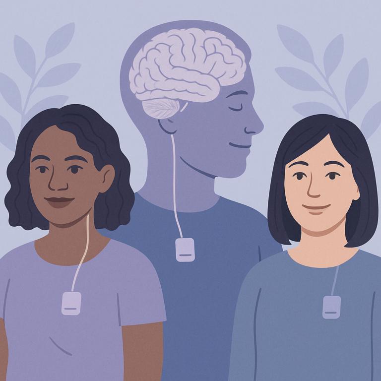
Researchers studied juvenile absence epilepsy (JAE) to understand how it affects the brain and cognitive abilities. They looked at 23 individuals with JAE, 18 of their unaffected siblings, and 28 people without any epilepsy. The study used advanced brain imaging techniques to see how the brains of these groups functioned and were structured, especially during tasks that required working memory.
The key findings showed that individuals with JAE had different brain activity compared to their siblings and controls. They had increased activity in the motor cortex when focusing on attention tasks but showed less motor activity when working memory demands increased. Additionally, certain areas of the brain, particularly in the frontal regions, had reduced gray matter volume in JAE patients, which was linked to language difficulties. Interestingly, both JAE patients and their siblings had increased gray matter in another brain area, suggesting a possible shared trait.
These findings are important because they highlight how JAE affects the brain differently than other types of epilepsy, like juvenile myoclonic epilepsy (JME). Understanding these differences can help in tailoring treatments and support for individuals with JAE. However, the study had a small number of participants, so more research is needed to confirm these results and explore their implications further.
Free: Seizure First Aid Quick Guide (PDF)
Plus one plain-language weekly digest of new epilepsy research.
Unsubscribe anytime. No medical advice.