EEG in Epilepsy: Brain Signals, Studies, and Context
A plain-language guide to scalp EEG, newer signal measures, and monitoring studies for families who keep seeing “biomarker” and “brain waves” in the news.
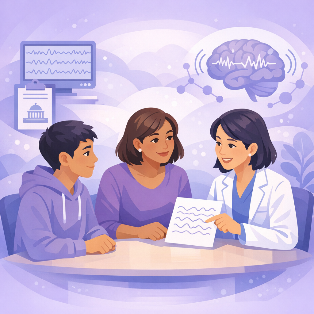
Neurology teams use EEG and other brain-signal tools to answer different questions at different times. They may use them during diagnosis, medication changes, hospital monitoring, and sometimes surgery planning. Research summaries on this site often use terms like biomarker, spikes, connectivity, and stereo-EEG. This hub links those ideas in one place.
How this hub helps
For the full, newest-first list of posts in this area, visit the Imaging and EEG topic archive. This hub is the guided tour. The archive is the shelf.
Some summaries focus on surgery planning, invasive EEG, or neuromodulation. Those topics still involve brain signals, but they raise different decisions. Ask your care team how any study applies to you or your child.
EEG in the headlines
EEG measures electrical activity from the brain’s surface. In specialized settings, teams may also record signals with electrodes placed for careful mapping. Many studies look for patterns that may predict outcomes, not brand-new gadgets for home use.
What biomarker means
When you read biomarker, think measurable clue, not automatic diagnosis. Strong articles explain who researchers studied, what they measured, and what still needs to happen before clinics use it widely.
Ask your neurologist
Bring questions if a headline suggests a test will pick the perfect drug, replace monitoring, or predict surgery success by itself. Those stories are often early steps, not final answers.
What this means for you
- In everyday terms, a biomarker is something measurable that helps clinicians sort risk, choose a next test, or compare treatments. In epilepsy research, EEG patterns and related signals often fill that role, even when they cannot guide care on their own yet.
- Scalp EEG, short or long monitoring, and research algorithms all have limits. A finding may be real in a study and still not give you something useful to act on at home.
- Posts that mention sEEG, stereo-EEG, or grids usually connect most closely to surgical evaluation. Read them as background on how centers map seizures, not as a reason to change your medical plan without your team.
Three paths families often take
Making sense of research on brain signals
Start here when a summary promises smarter tools, better prediction, or “new insights” from EEG data. These pieces explain what was actually measured and what is still experimental.
- New insights on brain signals for epilepsy treatment
- EEG abnormalities and language delays in children
- Using EEG data to localize seizure-related brain areas
When epilepsy surgery or advanced mapping comes up
Invasive EEG and connectivity studies usually sit inside a specialized center evaluation. Read them for context on how teams plan safe, precise treatment, not as instructions to pursue surgery on your own.
- Brain connectivity and epilepsy surgery
- Improving accuracy in stereoelectroencephalography (sEEG)
- iEEG connectivity and surgery outcomes
Monitoring, prognosis, and hospital EEG stories
Longer monitoring and ICU-style EEG show up in very different settings from a routine office visit. These summaries help you separate “interesting signal science” from what your team might order next.
- Seizure clusters in epilepsy monitoring units
- Predicting recovery after cardiac arrest using EEG patterns
- Wearable devices and seizure detection research
EEG and related tests at a glance
Routine scalp EEG Often used to capture brain wave patterns during a short visit. Useful for diagnosis and some treatment checks, but may miss events that happen rarely or only during sleep.
Ambulatory or longer outpatient monitoring Extends recording time at home or over days to catch spells that a short study might miss. Helpful for symptoms that do not line up with a single office visit.
Continuous EEG in the hospital (cEEG) Used when teams need longer monitoring around the clock, for example, when someone is very ill, recovering from a brain injury, or when subtle changes need close observation. Not the same setting as a routine clinic appointment.
Invasive monitoring (e.g., sEEG) Electrodes placed during a careful epilepsy center workup to map seizures when surgery or targeted therapy is on the table. Always specialized, team-driven, and not implied by a routine EEG alone.
Safety and expectations
Research headlines move faster than clinic guidelines. Use what you read to prepare questions, not to start, stop, or change medications or devices without your clinician.
If a study mentions machine learning, connectivity scores, or wearable detection, ask what population it studied and whether your hospital offers anything similar today.
Posts about ICU EEG or cardiac arrest prognosis describe different patient groups from most epilepsy clinic visits. Notice the setting before you compare the story to your own experience.
Each linked summary keeps its original publication date. Newer articles are not “more correct” by default, check what the study design actually supports.
Quick answers
Timeline
Before your visit Read a few summaries that match your situation, such as pediatric EEG, long-term monitoring, or posts about prognosis. Write down terms you do not know, and note the date of each article.
During the conversation Ask how a pattern or score from a study fits your records, whether repeat testing is needed, and what would change based on the result. Also ask what is still experimental.
After you read news at home If a summary brings up hope or fear, check whether it came from a small cohort, a single center, or animal work. Share the link with your team instead of changing medicines or devices on your own.
Glossary
Related research on Epilepsy Explained
Featured summaries
SEEG and SDE Are Safe Options for Seizure Localization – Epilepsy Explained
Discontinuing Antiseizure Medications Early Is Safe for Infants – Epilepsy Explained
New Insights on Managing Seizure Clusters in Epilepsy Monitoring Units – Epilepsy Explained
More research
- Epilepsy Imaging & EEG: Explained | Epilepsy Explained
- Seizure Risks Linked to Insomnia Medications in Epilepsy Patients – Epilepsy Explained
- EEG Abnormalities Common in Children with Language Delays – Epilepsy Explained
- New Insights on Brain Signals for Better Epilepsy Treatment – Epilepsy Explained
- Understanding Brain Connectivity in Epilepsy Surgery – Epilepsy Explained
- New-Onset Seizures Linked to COVID-19 Show Good Prognosis – Epilepsy Explained
- Predicting Recovery After Cardiac Arrest Using EEG Patterns – Epilepsy Explained
- Longer Epilepsy Duration Increases Brain Connectivity in Children – Epilepsy Explained
- New Method Identifies Seizure Areas Using EEG Data – Epilepsy Explained
For the full feed of posts on this topic, see all Imaging and EEG articles.