Drug-Resistant Epilepsy | Epilepsy Research Summaries | Genetics | Imaging & EEG | New Studies | Pediatrics
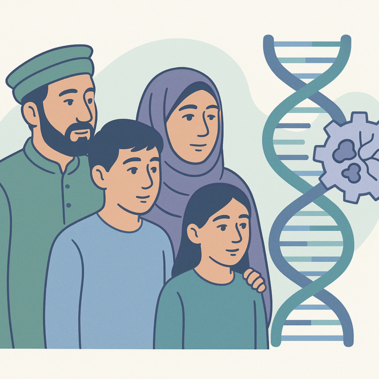
New Genetic Variants Found in Epilepsy Among Pashtun Families
This study focused on families from the Pashtun population in Pakistan who have cases of epilepsy that had not been thoroughly examined at the genetic level.